

|
Getting your Trinity Audio player ready...
|
✓ Medically reviewed by Dr. Ashutosh Shah, Plastic, Reconstructive & Cosmetic Surgeon (M.Ch., DNB)
Breast tissue testing after reduction helps doctors check removed breast tissue for hidden abnormalities. A pathologist examines tissue samples under a microscope through a process called histopathology. This examination can reveal normal tissue, benign changes, unusual cells or, rarely, unexpected cancer. Most reports show benign findings. However, the results help doctors decide whether a patient needs additional tests or follow-up care.
Breast reduction surgery, also called reduction mammoplasty, removes excess breast tissue, fat and skin. As a result, many patients experience improved comfort and a smaller breast size.
Women often choose this surgery to address back pain, shoulder discomfort, skin irritation or difficulty wearing supportive clothing.
However, patients may wonder what happens to the breast tissue after surgery.
Does the hospital discard it? Does a laboratory examine it? Can doctors discover an unexpected breast condition?
Many hospitals routinely send removed tissue to a pathology laboratory. Therefore, understanding this process can help patients feel more confident about their postoperative care.
Why Is Breast Tissue Testing After Reduction Important?
Breast tissue testing after reduction helps doctors identify microscopic changes that a routine examination may miss.
Breast tissue contains milk ducts, glands, connective tissue and fat. Sometimes, abnormal cells develop within these structures without causing noticeable symptoms.
For example, a woman may have no breast lump or pain. Nevertheless, a pathologist may discover a small abnormality while examining tissue samples.
Histopathology can identify several findings:
- Normal breast tissue
- Fibrocystic changes
- Benign cysts
- Fibroadenoma
- Usual ductal hyperplasia
- Atypical ductal or lobular hyperplasia
- Ductal carcinoma in situ (DCIS)
- Rarely, invasive breast cancer
Fortunately, most breast reduction specimens show benign changes.
However, an unexpected finding may influence future screening or treatment decisions.
Can Normal-Looking Breast Tissue Contain Abnormal Cells?
Yes.
Some abnormalities are too small for surgeons to recognise during an operation.
Additionally, a patient may have no symptoms despite microscopic changes in the breast.
A pathologist examines selected tissue sections under a microscope to identify these changes.
However, routine sampling cannot check every cell. Therefore, patients should continue appropriate breast screening even after receiving a benign report.
What Is Histopathology After Breast Reduction?
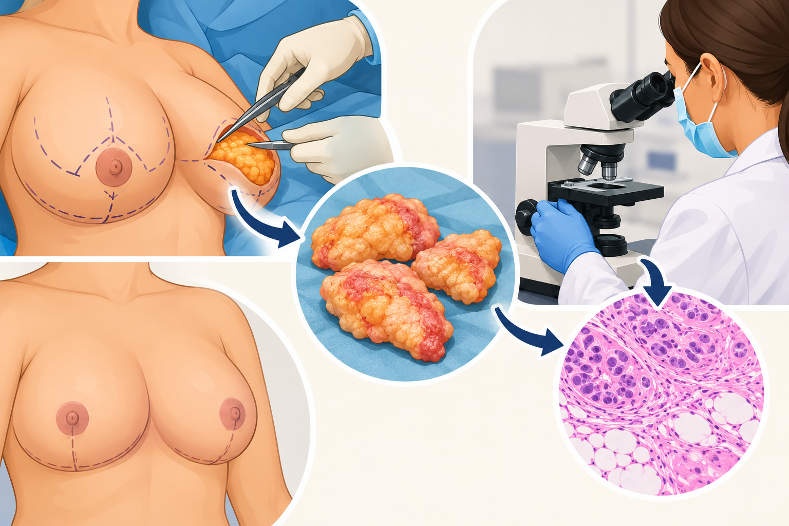
Histopathology after breast reduction is a laboratory process that examines removed breast tissue.
A pathologist studies the tissue to identify normal structures and possible abnormalities.
The examination usually follows several steps.
Step 1: Collecting Breast Tissue
First, the surgeon removes the planned amount of breast tissue during reduction surgery.
The surgical team then collects the tissue for laboratory examination.
Step 2: Identifying the Specimen
Next, the team places the tissue in a suitable container and adds the necessary identification details.
The team generally labels right and left breast specimens separately.
This process helps the laboratory identify the correct source of each sample.
Step 3: Examining the Tissue
At the laboratory, a pathologist or trained professional examines the specimen.
They measure, weigh and inspect the tissue for visible changes.
Step 4: Selecting Tissue Samples
The laboratory team selects representative tissue sections.
If they notice a suspicious area, they may collect additional samples.
Step 5: Preparing Microscope Slides
Next, laboratory professionals process the samples and prepare thin tissue sections.
They use special stains to make cellular structures easier to identify.
Step 6: Studying the Slides
Finally, the pathologist examines the slides under a microscope.
They document the findings in a histopathology report.
During breast tissue testing after reduction, the pathologist may request additional sections or tests if the initial findings need clarification.
For further information, read the National Cancer Institute’s guide to surgical pathology reports.
What Can Breast Tissue Testing After Reduction Detect?
A breast tissue examination can reveal several conditions.
However, an abnormal result does not always mean cancer.
Many findings are harmless and require no specific treatment. In contrast, some changes may increase future breast cancer risk.
Common Histopathology Findings
| Finding | What It Means | Possible Next Step |
|---|---|---|
| Normal breast tissue | No significant abnormality appears in the examined sections | Continue routine screening |
| Fibrocystic changes | Common benign changes affect breast glands and connective tissue | Usually no treatment |
| Fibroadenoma | A non-cancerous breast tumour | Clinical review if necessary |
| Usual ductal hyperplasia | Breast ducts contain extra non-atypical cells | Follow risk-based screening |
| Atypical hyperplasia | Certain abnormal cells indicate increased future cancer risk | Consult a breast specialist |
| DCIS | Cancer cells remain within breast ducts | Seek specialist evaluation |
| Invasive carcinoma | Cancer cells extend beyond the original duct or lobule | Arrange prompt specialist care |
The next step depends on the exact diagnosis, medical history and imaging findings.
Are Fibrocystic Changes Dangerous?
Most fibrocystic changes are benign.
They may involve small cysts, fibrosis or other non-cancerous changes.
Many women experience these changes without noticeable symptoms.
Therefore, a pathology report helps doctors distinguish common benign findings from conditions that need further assessment.
What Is Atypical Hyperplasia?
Atypical hyperplasia involves unusual cell growth within breast ducts or lobules.
It is not invasive breast cancer.
However, certain types increase future breast cancer risk.
Consequently, a doctor may recommend a specialist consultation, personalised screening or a discussion about risk-reduction options.
Can Breast Cancer Be Found During Breast Reduction Surgery?
Yes, although unexpected cancer findings are uncommon.
Sometimes, a pathologist discovers a high-risk lesion or cancer in tissue removed during breast reduction.
Doctors call this an incidental pathological finding.
The likelihood varies according to age, medical history, breast cancer risk factors and screening practices.
Why Might Doctors Miss Cancer Before Surgery?
Some breast abnormalities are extremely small.
Others cause no symptoms or visible changes.
Therefore, a lesion may remain undetected until the pathologist examines the removed tissue.
However, histopathology does not replace appropriate breast screening before surgery.
Is Breast Imaging Necessary Before Reduction Surgery?
The surgeon considers the patient’s age, symptoms, family history and individual risk factors.
Depending on these factors, the patient may need mammography, ultrasound or additional assessment.
Patients should report symptoms such as:
- A new breast lump
- Unexplained nipple discharge
- Persistent pain in one area
- Unusual skin changes
- New nipple inversion
- Previous abnormal imaging results
If the surgeon identifies a concern, further assessment may be necessary before surgery.
Is Breast Tissue Testing Necessary for Every Patient?
Hospitals and healthcare systems follow different specimen-testing policies.
Many hospitals routinely examine breast reduction tissue. However, other facilities follow protocols that consider patient risk and local guidelines.
Because microscopic abnormalities can occur without symptoms, many surgical teams favour routine examination.
Patients should therefore ask their surgeon about the hospital’s testing policy.
Does Age Affect the Need for Testing?
Breast cancer risk generally increases with age.
However, younger women can also develop benign or atypical breast conditions.
Doctors consider several factors:
- Patient age
- Family history
- Previous breast abnormalities
- Personal history of breast disease
- Known inherited cancer-risk variants
- Previous abnormal imaging
- New breast symptoms
Together, these details help the surgeon plan appropriate care.
How Long Does Histopathology After Breast Reduction Take?
Many laboratories provide routine histopathology reports within several working days to approximately one or two weeks.
However, turnaround times vary.
Some specimens require additional examination. As a result, the laboratory may need more time.
Why Can a Report Take Longer?
Several factors may affect the timeline:
- Large tissue specimens
- Additional microscopic sections
- Unusual findings
- Special staining procedures
- Specialist pathology review
- Laboratory workload
Importantly, a delayed report does not automatically indicate cancer.
Patients should ask their surgeon when to expect the results.
How Do You Read a Breast Reduction Histopathology Report?
A pathology report contains several sections.
Although the terminology may seem complex, understanding the basic structure can help patients discuss the results with their doctor.
1. Specimen Details
This section identifies the tissue that the laboratory received.
For example, it may specify the right or left breast.
2. Gross Description
Here, the laboratory describes the specimen’s weight, size and visible characteristics.
3. Microscopic Description
The pathologist records the changes observed under the microscope.
4. Final Diagnosis
This section summarises the main findings.
For example, the report may describe normal tissue, benign changes or a specific abnormality.
5. Additional Comments
Sometimes, the pathologist recommends further investigation or comparison with imaging findings.
The surgeon should explain the results of breast tissue testing after reduction and clarify whether the patient needs additional care.
What Happens if Abnormal Cells Are Found?
The next step depends on the type of abnormality.
For example, common benign changes may require no treatment.
However, atypical hyperplasia may require specialist risk assessment.
If the report identifies cancer, the surgeon should arrange prompt evaluation by a breast cancer specialist.
What Additional Tests Might Doctors Recommend?
Depending on the findings, a specialist may recommend:
- Diagnostic mammography
- Breast ultrasound
- MRI in selected cases
- Pathology slide review
- Additional biopsy
- Assessment of remaining breast tissue
- Individualised treatment planning
Importantly, breast reduction surgery differs from planned cancer surgery.
During routine reduction, the surgical team may not map the tissue in the same way as a cancer operation.
Therefore, an unexpected cancer finding can make it difficult to identify the original lesion location.
A breast specialist can assess the situation and recommend the next steps.
Does Histopathology Affect Breast Reduction Recovery?
No. The laboratory examination itself does not affect physical recovery.
The pathologist examines tissue that the surgeon has already removed.
Therefore, routine testing does not require an additional procedure.
After breast reduction, patients may experience:
- Temporary swelling
- Bruising
- Wound discomfort
- Healing incisions
- Temporary activity restrictions
- A need for supportive garments
Meanwhile, the laboratory examines the tissue.
If the report identifies an important abnormality, the surgeon may arrange further appointments.
Should Patients Wait for the Report Before Resuming Activities?
Not usually.
The surgeon bases routine activity advice on wound healing, the extent of surgery and individual recovery.
However, patients should follow all postoperative instructions.
If the report identifies a significant finding, additional medical appointments may become necessary.
Does Breast Tissue Examination Cost Extra?
The cost depends on the hospital, laboratory and surgical package.
Some hospitals include routine histopathology in their surgical quotation.
Others charge separately.
Additionally, special stains or specialist reviews may increase the total cost.
Before surgery, ask these questions:
- Does the quotation include histopathology?
- Will the laboratory examine both breast specimens?
- Are additional tests charged separately?
- When can I expect the report?
- Who will explain the findings?
- What happens if the report identifies an abnormality?
These questions help patients understand the process and avoid unexpected expenses.
Why Reviewing the Pathology Report Matters
After breast reduction, patients naturally focus on comfort, breast shape and wound healing.
However, reviewing the histopathology report is also important.
Even when the patient feels well, the surgeon should review the results and explain any significant findings.
At Elegance Clinic, Dr. Ashutosh A Shah, MBBS, MS, MCh and DNB, Plastic and Reconstructive Surgeon with over 22 years of experience, discusses breast reduction planning, recovery and postoperative pathology review with patients. If the report identifies a high-risk or malignant finding, appropriate specialist assessment and follow-up become important.
For additional information, explore breast treatment services.
Frequently Asked Questions
1. Is Removed Breast Tissue Always Sent for Testing?
Many hospitals routinely send removed breast tissue to a pathology laboratory. However, policies vary. Therefore, patients should confirm the testing procedure with their surgeon.
2. Can Breast Cancer Be Found During Breast Reduction?
Yes. Occasionally, a pathologist discovers an unexpected cancer or high-risk lesion. Fortunately, most breast reduction specimens show benign findings.
3. How Long Does a Breast Tissue Histopathology Report Take?
Many laboratories provide routine reports within several working days to one or two weeks. However, additional testing may take longer.
4. What Does a Benign Breast Tissue Report Mean?
A benign report means the pathologist found no malignant changes in the tissue sections examined. Nevertheless, patients should continue appropriate breast screening.
5. What Happens if Abnormal Cells Are Detected?
The next step depends on the diagnosis. Some findings require only routine care. However, atypical changes or cancer may require specialist evaluation.
6. Does Breast Tissue Testing Cost Extra?
Some hospitals include testing in the surgical package, while others charge separately. Therefore, patients should check the quotation before surgery.
7. Is Breast Tissue Testing Painful?
No. The laboratory examines tissue that the surgeon has already removed. As a result, the examination itself causes no additional pain.
8. Can Histopathology Detect Every Breast Abnormality?
No. Pathologists examine selected tissue sections rather than every cell. Therefore, routine screening and medical assessment remain important.
Conclusion
Breast tissue testing after reduction helps doctors identify important changes in removed breast tissue.
In most cases, the pathologist finds normal or benign changes.
However, testing may occasionally reveal atypical cells or unexpected cancer.
Therefore, patients should ask when the report will arrive and who will explain the results.
A timely review helps the surgical team identify findings that require further care.
Ultimately, breast reduction care involves more than improving breast size and comfort. Appropriate tissue examination and follow-up also support informed long-term breast health decisions.
